 Massage & Bodywork magazine is a bimonthly journal for a national audience of massage, bodywork, and somatic professionals. Each issue of our award-winning massage magazine (included as part of ABMP membership, which also includes professional liability insurance) offers in-depth articles on subjects important to the practicing professional and the layperson. The latest research, historical perspectives, massage techniques, business information, professional trends, and a variety of regularly featured columns keep Massage & Bodywork readers in touch with their work, their clients, and their health.
ABMP members receive the print version of Massage & Bodywork six times per year, and the digital edition is available online at www.massageandbodyworkdigital.com.
Here’s what Massage & Bodywork readers say:
- “Massage & Bodywork is an invaluable tool for me on many aspects of our profession.”
- “I find the articles very enlightening and conducive to the continuation of my education.”
- “Thank you so much for a professional and informative magazine for massage therapists.”
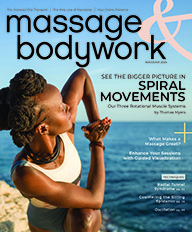
Read the current Massage & Bodywork issue.
Visit the Massage & Bodywork digital archive to access even more insider information and industry updates for professional massage therapists.
Blind and visually impaired readers, or those wishing to read the content in a text-only format, are able to access readable files with our Text-Only Magazines. These files are compatible with JAWS, Window Eyes, and Zoom Text.
Want to work with Massage & Bodywork magazine?
Submit an article for consideration to Massage & Bodywork magazine.
Learn about how to advertise in Massage & Bodywork magazine by contacting our advertising department at advertising@abmp.com.